Kapitel 1
The Invisible Force That Shapes Our World
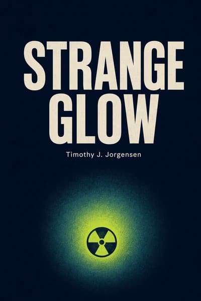
In a world increasingly concerned with invisible threats, few subjects generate more visceral fear than radiation. Timothy J. Jorgensen's "Strange Glow" cuts through this fear with scientific clarity and historical richness. This book has become required reading in university physics and public health programs worldwide, praised by Nobel laureates for its exceptional ability to make complex radiation science accessible. Even Elon Musk reportedly keeps a copy on his nightstand, curious about the force that both powers spacecraft and poses existential risks. What makes this book revolutionary is how it transforms our relationship with radiation from one of blind fear to informed respect-much like humanity's journey with electricity a century ago.
Kapitel 2
Radiation: The Misunderstood Phenomenon
Fear distorts our perception of radiation in profound ways. The word instantly conjures images of mushroom clouds and cancerous tumors, creating an anxiety that's both understandable and potentially harmful. While some radiation sources warrant legitimate concern, many people have developed excessive fear of diagnostic x-rays, airport scanners, cell phones, and microwave ovens-an anxiety level that's unwarranted and sometimes dangerous.
Research shows that fear hijacks our risk assessment abilities. We tend to overestimate risks from things we fear while underestimating risks from less frightening hazards. Consider how many Americans fear black widow spiders despite fewer than two annual deaths, while ignoring mosquitoes that cause over 1,000 serious illnesses and deaths yearly. Similarly, some motorcyclists fear flying despite the vastly higher risk of motorcycle crashes.
This pattern repeats with radiation. When irrational fear takes control, we struggle to weigh risks objectively and may make choices that actually increase rather than decrease our personal risk. Understanding radiation's basic principles helps us develop a more balanced perspective.
Radiation is simply energy in motion, traveling through matter or space. While most radiation is invisible, light is the one type we can see with our naked eyes. By understanding light's visible properties, we gain insight into all radiation forms. Throughout history, humans correctly deduced that the sun was a massive ball of fire, noting that heat and light, while often occurring together, are separate phenomena.
The difference between various forms of electromagnetic radiation lies in their wavelengths. X-rays have extremely short wavelengths (less than a human hair's width), while radio waves can span the length of a football field. Shorter wavelengths deliver more energy-similar to how closely spaced ocean waves hit a beach with greater frequency and force. This wavelength difference determines radiation's properties, including penetration ability and potential health effects.
Kapitel 3
The Accidental Discovery That Changed Medicine
Wilhelm Conrad Roentgen's discovery of x-rays in 1895 was a true scientific accident. While experimenting with a Crookes tube (a vacuum tube with electrodes) in his darkened laboratory, he noticed that a nearby fluorescent screen would mysteriously glow whenever he ran current through the tube. These invisible rays could penetrate solid objects-passing through cardboard, books, and even human flesh while casting shadows of denser materials like bones and metal.
After photographing his wife's ringed hand-producing the first x-ray image that both amazed and terrified her ("I have seen my own death!")-Roentgen published his findings in December 1895. News spread worldwide within days, and within weeks his results were replicated at Harvard, Dartmouth, and Princeton. Just six weeks after Roentgen's discovery, doctors in Montreal used x-rays to locate and remove a bullet in patient Toulson Cunning's leg-the fastest bench-to-bedside application of any scientific discovery in history.
Unlike many scientists of his era, Roentgen never patented his discovery, considering himself a pure scientist uninterested in practical applications. This decision accelerated x-ray technology's adoption worldwide but cost him potential fortune. For his contribution, Roentgen received the first Nobel Prize in Physics in 1901.
The medical community immediately recognized x-rays' diagnostic potential, but another application emerged just as quickly. In January 1896-just one month after Roentgen's publication-20-year-old Emil Herman Grubbe began treating cancer patients with x-rays. His first patient, Mrs. Rose Lee with advanced breast cancer, received 18 treatments that reduced her pain before she died a month later. Though initially sent only terminal cases, Grubbe persisted until doctors began referring patients with earlier-stage disease who showed remarkable improvement.
Grubbe's approach of fractionating radiation doses-delivering small amounts over multiple sessions-proved crucial to his success, though he didn't understand why. Scientists later discovered that rapidly dividing cells (like tumor cells) are more sensitive to radiation than slower-growing normal cells, and this differential sensitivity is enhanced through dose fractionation.
Kapitel 4
Radioactivity: Nature's Hidden Energy
While Roentgen discovered x-rays produced by machines, Antoine Henri Becquerel accidentally discovered natural radioactivity in 1896. Following in his family's tradition of studying fluorescence, Becquerel was testing whether uranium compounds emitted x-rays when exposed to sunlight. To his surprise, he found that uranium exposed photographic film regardless of light exposure. Becquerel realized uranium atoms spontaneously emitted invisible penetrating radiation unrelated to fluorescence.
This discovery revealed that radioactivity is an atom's intrinsic ability to release radiation without external stimulation. While uranium was the first radioactive substance discovered, hundreds of radioisotopes exist, many mixed with their non-radioactive forms. Some radioactive elements are essential to life, like carbon and potassium, making all living things naturally radioactive to some degree.
Marie and Pierre Curie expanded on Becquerel's work, processing tons of pitchblende ore to isolate just 0.1 gram of radium. Their work revealed two previously unknown elements-polonium (named for Marie's native Poland) and radium (from the Latin for "ray"). While Becquerel merely discovered radioactivity in uranium, the Curies actually added new elements to the periodic table.
Atomic stability depends on the balance between protons and neutrons in a nucleus. Stable atoms typically have a nearly one-to-one ratio of these particles. When this balance is disrupted, the nucleus becomes unstable and undergoes radioactive decay to correct the imbalance. For example, carbon-14 (with six protons and eight neutrons) is unstable and decays by converting a neutron into a proton, becoming nitrogen-14 (with seven protons and seven neutrons).
The half-life concept reveals how unstable a radioisotope is-an intrinsic property that cannot be altered. Carbon-14's half-life of 5,730 years means one gram would become 0.5 gram in that time, with the remainder transformed to nitrogen-14. This predictable decay enables radiocarbon dating of ancient biological artifacts, as the carbon-14 to carbon-12 ratio decreases at a known rate after death.
Kapitel 5
The Atomic Revolution
Ernest Rutherford revolutionized our understanding of atomic structure through his famous gold foil experiment in 1909. Using alpha particles (essentially helium nuclei) as atomic probes, he discovered that most particles passed through the foil but some bounced back-an event he described as "incredible as if you fired a 15-inch shell at tissue paper and it came back to hit you." This unexpected result shattered the prevailing "plum pudding" model of the atom, which had suggested that electrons were embedded throughout a positive mass. Instead, Rutherford proposed a new planetary model with a tiny, dense nucleus surrounded by mostly empty space where electrons orbited, similar to planets around the sun.
The discovery raised new questions about atomic structure. Rutherford proposed the existence of neutrons-particles with the same mass as protons but no charge-to solve a fundamental puzzle: how atoms could contain their observed mass without having excessive positive charge that would tear them apart. His colleague James Chadwick spent years pursuing this hypothesis, finally confirming the existence of neutrons in 1932 through experiments with beryllium radiation. This completed the basic model of atomic structure: protons and neutrons constituting the nucleus's mass with electrons orbiting in the surrounding space, held in place by electromagnetic forces.
While Chadwick focused on neutrons, other Cavendish scientists John Cockcroft and Ernest Walton pursued an even more ambitious goal-artificially splitting the atom. They designed and built a remarkable 700,000-volt particle accelerator, an achievement in itself given the technical limitations of the 1930s. Their persistence paid off when they successfully split lithium-7 atoms, producing visible alpha particle tracks when bombarded with protons. This historic achievement in 1932 earned them the Nobel Prize and opened the door to controlled nuclear reactions.
Nuclear reactions presented scientists with a perplexing problem: careful measurements showed that the products consistently weighed slightly less than the reactants, seemingly violating the fundamental law of conservation of mass. Einstein's famous equation E=mc2 provided the elegant solution by demonstrating that mass and energy are interchangeable forms of the same thing. When an atom undergoes decay or fission, it releases energy equivalent to the mass lost, following the precise relationship Einstein described. The implications of this mass-energy equivalence are staggering-just one gram of matter completely converted to energy would yield 90 trillion joules, equivalent to 10,000 lightning bolts or one atomic bomb. This understanding laid the groundwork for both peaceful nuclear power generation and the development of nuclear weapons, fundamentally changing human society's relationship with energy and warfare.
The atomic revolution of the early 20th century transformed not just physics but our entire understanding of matter, energy, and the universe itself. These discoveries led directly to technologies that define the modern world, from nuclear medicine to power plants, and continue to influence scientific research today.
Kapitel 6
The Deadly Price of Progress
While scientists explored radiation's fundamental properties, workers in various industries were unwittingly exposing themselves to dangerous levels. The tragic story of the "radium girls"-young women who painted watch dials with radium-laced paint in the 1920s-illustrates radiation's hidden dangers.
These women were instructed to point their paintbrushes with their lips, ingesting small amounts of radium with each lick. Unlike the Schneeberg miners who suffered lung ailments from inhaled radon, the dial painters experienced bone problems because their ingested radium settled in their skeletons. Radium's chemical similarity to calcium caused it to be incorporated into bone where it continuously irradiated surrounding living tissues.
Frances Splettstocher was just one of many young women who suffered horrific deaths. After years of denial, watch companies finally accepted blame and paid settlements totaling $90,000 ($1.5 million in 2015 dollars) to 16 affected women. By 1927, safety measures were implemented-fume hoods, hairnets, gloves, and a ban on lip-pointing brushes-which effectively prevented health problems in new workers.
Even the wealthy weren't immune to radiation's dangers. Industrialist and golf champion Eben McBurney Byers began drinking Radithor-radium dissolved in distilled water-in 1927 to heal a broken arm. He consumed approximately 1,400 bottles over three years, losing most of his jaw and developing holes in his skull before dying in 1932.
Marie Curie herself developed serious health problems including cataracts and eventually died of extreme anemia in 1937, her bone marrow permanently damaged from accumulated radiation exposure. Contrary to popular belief, her 1995 exhumation revealed she hadn't died from internal radium poisoning but rather from external gamma and x-ray exposures.
Kapitel 7
Understanding Radiation Sickness
The atomic bombings of Hiroshima and Nagasaki provided tragic but scientifically valuable insights into radiation's health effects. Dr. Terufumi Sasaki, a 25-year-old physician at Hiroshima's Red Cross Hospital, was one of the few medical professionals who escaped serious injury. Of the hospital's 30 doctors, only 6 could function; of 200 nurses, only 10 could work. This small team faced 10,000 bomb victims descending upon their 600-bed hospital.
Sasaki and his staff recognized that radiation sickness manifested in three successive waves, each affecting different groups of victims who had received different radiation doses:
The first wave struck those closest to the blast who received the highest doses (over 20,000 mSv). These victims suffered the Central Nervous System syndrome as radiation killed even their brain neurons, causing brain swelling, coma, and death within three days.
The second wave arrived about a week later, affecting victims with severe gastrointestinal problems, hair loss, and high fever. These patients had received doses between 10,000-20,000 mSv, enough to kill the rapidly dividing crypt cells in their intestines. None survived beyond two weeks after the bombing.
After another lull of several weeks, a third wave of deaths began around thirty days after the bombing. These patients experienced complete loss of all blood cell types (pancytopenia) and died from related complications. This condition is called hematopoietic syndrome, occurring with whole-body doses of 1,000-10,000 mSv.
By November 1945, Hiroshima's radiation syndromes had run their course. Those destined to die had done so, while survivors were recovering, though many experienced lingering side effects like chronic fatigue. Death toll estimates from the Hiroshima bombing range from 90,000 to 165,000, with approximately 75% dying from fire and trauma while 25% succumbed to radiation effects.
Kapitel 8
Radiation's Long-Term Legacy
While acute radiation sickness claimed many lives immediately after exposure, scientists soon recognized that survivors faced increased cancer risks years later. Japanese atomic bomb survivors feared purple spots (petechiae) on their skin as harbingers of leukemia. Dr. Fumio Shigeto noticed elevated leukemia rates among survivors by 1948, meticulously documenting cases despite initial skepticism from Japanese and American officials.
By 1953, leukemia rates peaked and began subsiding, but starting in 1955, many other cancer types appeared, continuing to rise until 1982. Unlike chemical carcinogens that target specific tissues, radiation proved to be an unselective carcinogen affecting virtually all body tissues.
The atomic bomb survivors represented an unprecedented scientific opportunity to measure radiation's cancer risks precisely. They were "the most important people living" according to radiation scientist Dr. Robert H. Holmes, because they offered two crucial advantages: a large population of all ages and sexes exposed to various radiation doses simultaneously, and the ability to determine individual doses based on each person's exact location during the bombing.
President Harry Truman established the Atomic Bomb Casualty Commission in 1946, which launched the Life Span Study (LSS) tracking 120,000 survivors and controls for over 65 years-now considered the definitive epidemiological study on radiation's health effects.
The LSS has yielded a precise cancer risk estimate: 0.005% increased lifetime cancer risk per millisievert of whole-body radiation. This allows us to convert any radiation exposure into a meaningful risk assessment. For example, a whole-body spiral CT scan delivering 20 mSv represents a 0.1% increased lifetime cancer risk-equivalent to 1 in 1,000 people developing cancer from such a scan, or increasing one's baseline cancer risk from approximately 25% to 25.1%.
Kapitel 9
DNA: The Target for Radiation Damage
Scientists eventually discovered that radiation's biological effects result from damage to DNA-the genetic material in our cells. Through meticulous experimentation in the 1920s and 1930s, radiation biologists confirmed that to kill a cell with radiation, its nucleus must be irradiated, and cells could be rescued by transplanting non-irradiated nuclei. Microscopic examination revealed radiation breaks chromosomes in distinctive patterns, with biological effects directly proportional to the extent of chromosome damage. These breakthrough experiments, particularly those conducted by Hermann Muller using fruit flies, demonstrated that radiation could induce heritable mutations.
With DNA established as the substance of genes through the work of Avery, MacLeod, and McCarty in 1944, the conclusion became clear: DNA is radiation's primary biological target. This doesn't mean radiation specifically aims for DNA, but rather that DNA damage is necessary and sufficient for significant biological effects. While radiation indiscriminately damages many cellular molecules, including proteins, lipids, and carbohydrates, only DNA damage produces lasting consequences because other molecules are readily replaceable under DNA's direction. When DNA-the cellular director-is damaged, proper replacement becomes impossible, resulting in either cell death or mutations that can lead to inheritable defects or cancer.
The discovery of DNA's double helix structure by James Watson and Francis Crick in 1953 revolutionized our understanding of how radiation damages genes at the molecular level. Their model revealed that DNA consists of two complementary strands twisted together, with the precise sequence of nucleotide bases carrying genetic information. Radiation can break these strands in several ways: causing single-strand breaks, double-strand breaks, base modifications, and cross-linking between DNA strands. These different types of damage require distinct repair mechanisms, with double-strand breaks being particularly dangerous because they're harder to repair accurately.
The relationship between radiation dose and DNA damage follows clear patterns. At high doses, multiple DNA breaks overwhelm cellular repair mechanisms, leading to cell death. At lower doses, fewer breaks occur, but if DNA repair systems fail to correct all damage, mutations can result. Modern research has revealed sophisticated DNA repair mechanisms, including base excision repair, nucleotide excision repair, and double-strand break repair through homologous recombination or non-homologous end joining.
Regardless of dose level, cellular DNA remains radiation's critical target-causing cell death at high doses and potentially harmful mutations at low doses when DNA repair systems fail to correct all damage. While reducing radiation exposure reduces risk, completely avoiding all radiation exposure means foregoing beneficial radiation technologies in medicine, research, and industry. This creates a complex risk-benefit calculation that must consider both the potential harm from radiation exposure and the substantial benefits of its controlled use. Understanding DNA as radiation's target has led to improved radiation protection standards and better therapeutic applications in cancer treatment.
Kapitel 10
Weighing Radiation Risks in Modern Life
Today, we face radiation exposure decisions in many contexts-from medical procedures to environmental concerns. Understanding the risk-benefit balance helps us make informed choices.
Radon gas in homes presents a significant but often overlooked radiation risk. When Stanley Watras triggered radiation alarms at the nuclear plant where he worked in 1984, investigation revealed his home contained radon levels 20 times higher than typical uranium mines. His house sat atop a "radon spring" on the uranium-rich Reading Prong geological formation. The EPA has determined that home radon concentration should not exceed 4 pCi/L, as lifetime residence in a home with this radon level creates a 6.2% lung cancer risk for smokers but only 0.73% for nonsmokers.
Medical imaging presents another common radiation exposure scenario. Modern arm X-rays deliver an effective dose of only 0.001 mSv, carrying a lifetime cancer risk of just 1 in 20 million-making the benefits overwhelmingly outweigh the minimal risk. However, procedures like whole-body CT scans deliver about 20 mSv-40 times that of a mammogram and 20,000 times that of an arm x-ray, with corresponding increases in cancer risk.
Cell phone radiation has generated significant public concern despite limited evidence of harm. Despite cell phone usage increasing from 0% in 1980 to 91% by 2013, brain cancer incidence in the United States has remained unchanged for 40 years. Unlike the clear correlation between cigarette smoking and lung cancer rates, we see no corresponding rise in brain tumors despite mobile devices now outnumbering people.
The Fukushima nuclear disaster in 2011 raised fears about radioactive contamination in food. Marine biologist Daniel Madigan discovered that Pacific bluefin tuna had carried cesium-134 and cesium-137 from Japan to California waters. However, the radiation doses from consuming these fish were minimal-about 0.000005% increased lifetime cancer risk, an insignificant addition to our baseline 25% cancer risk.
Kapitel 11
Living Wisely with Radiation
Radiation is neither inherently good nor bad-it's a natural force that humans have learned to harness for both beneficial and destructive purposes. The key to living wisely with radiation lies in understanding its properties, measuring its effects accurately, and weighing risks against benefits in each specific context. From background radiation in soil and cosmic rays to medical imaging and nuclear power, radiation surrounds us in various forms, each requiring different approaches to risk management.
For most medical procedures, the benefits of radiation far outweigh the risks when the procedure is appropriate for the clinical condition. A chest X-ray, for example, delivers about 0.1 millisieverts of radiation - equivalent to about 10 days of natural background radiation - while providing crucial diagnostic information. Even higher-dose procedures like CT scans (5-10 millisieverts) remain valuable diagnostic tools when clinically indicated. Medical professionals follow the ALARA principle (As Low As Reasonably Achievable) to minimize exposure while maintaining diagnostic quality.
For environmental exposures like radon, we can take reasonable precautions without excessive fear. Regular home testing, proper ventilation, and basement sealing can effectively reduce radon exposure. In areas with naturally high background radiation, such as parts of Kerala, India, or Ramsar, Iran, populations have lived for generations without demonstrable adverse effects, suggesting our bodies have some capacity to adapt to varying radiation levels.
Consumer technologies present varying degrees of radiation exposure. Cell phones emit non-ionizing radiation at levels well below international safety standards. Simple practices like using headsets, speakerphone options, or texting can further reduce exposure. Similarly, airport security scanners, microwave ovens, and other common devices are designed with multiple safety features and strict emission controls.
The story of radiation parallels our experience with electricity a century ago. Once feared as mysterious and deadly, electricity became an accepted part of daily life as we developed the knowledge and technology to use it safely. The early days of X-rays saw both remarkable medical breakthroughs and tragic accidents before proper safety protocols were established. Similarly, with proper understanding and respect, radiation need not be a source of irrational fear but rather a powerful tool that, when used wisely, can enhance our lives and health.
As we navigate an increasingly complex technological world, the ability to assess radiation risks accurately becomes not just a scientific skill but an essential component of modern scientific literacy. Understanding concepts like dose-response relationships, relative risk, and the distinction between ionizing and non-ionizing radiation helps inform better decision-making. By replacing fear with knowledge, we can make better decisions about radiation exposure in our personal lives and as a society, whether considering medical procedures, choosing where to live, or evaluating new technologies.